- DME Channel on YouTube
We develop a suitable microscope for you for each special application!
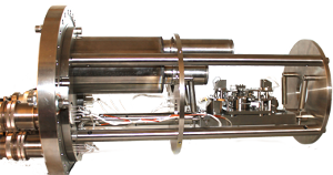
Highest precision UHV Scanning Tunneling Microscope is designed for 3-dimensional single atomic resolution and has the following properties:
Highest Performance:
|
Ease of use:
|
Flexibility:
|
Versatility:
|
Further information:
|
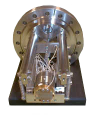
The DME Scanning Tunneling Microscope for Ultra High Vacuum is designed for 3-dimensional single atomic resolution and has the following properties:
Highest Performance:
|
Ease of use:
|
Flexibility:
|
Versatility:
|
Further information:
|
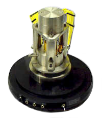
The Rasterscope™ ElectroChemical Scanning Tunneling Miscroscope (EC-STM) is dedicated for electrochemial investigation of surfaces in a liquid enviroment.
Highest Performance:
|
Ease of use:
|
Flexibility:
|
Versatility:
|
Further information:
|
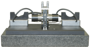
Using a head-to-head STM you can access the same point on both surfaces of a thin membrane simultaneously. This allows for example a direct measurement of a controlled deformation induced by one STM probe and monitored by the other STM.
First results on a few-layer graphene membrane can be read in the article Probing from Both Sides: Reshaping the Graphene Landscape via Face-to-Face Dual-Probe Microscopy by Jannik C. Meyer et al. (Department of Physics, University of Vienna) published in Nanoletters.
This system is a special version of a UHV Scanning Tunnelling Microscope allowing conducting tip enhanced raman spectroscopy (TERS) measurements.
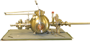
I.e. by nature, the signal to noise ratio in a Tip Enhanced Raman Spectroscopy (TERS) measurement is by nature very low, even
though the tip amplifies the Raman signal by many orders of magnitude. This is due to the high lateral resolution
and the low intensity of a Raman emission. To keep integration times for the optical measurement low,
the optical system must have a numerical aperture as high as possible.
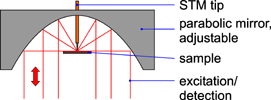 The sketch on the right shows a setup with a numerical aperture of nearly 1. Almost the whole half sphere
above the sample is used as light path.
- A 5 axis movable stage with a resolution of about 100 nm allows exact mirror positioning to perfectly align STM tip and focal point.
- Samples and STM tips can be exchanged without breaking the vacuum and without a risk of damage by a specially designed transfer mechanism.
- For vibrational damping purposes the whole optical and STM setup was designed on a free swinging platform. Light transfer is realized via glass fibers.
- The STM provides atomic resolution in all three dimensions.
With few modifications this system can also be equipped with a shear force AFM scanner, so that also non-conductive samples can be examined.
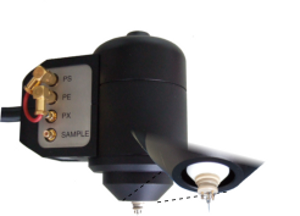
The tip scanner DS 95 is shear-force-microscope scanner, where the tip oscillates parallel to the sample surface. Instead of a cantilever, this scanner uses the sharpened end of an optical fiber as a scanning tip, which allows optical excitation and detection below the physical diffraction limit.
Depending on the type of SNOM experiment, many different measurement setups are possible, which can be realized by our manufactures.
The SNOM measurements done by the Institut für Angewandte Physik, Technische Universität Braunschweig show some examples of optical nearfield microscopy.
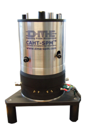
The controlled atmosphere high temperature AFM (CAHT) has been developed in order to perform electrochemical measurements on solid oxide fuel cells and electrolyzer cells, which can be used for electrochemical power conversion of gaseous fuel to electricity. The operation temperature of solid oxid fuel cells and electrolyzer is about 800°C at high surface temperatures.
The CAHT-SPM consists of two main parts: the upper detector part (metal) and the lower scanner part (black). The detector part is the cold part of the microscope, where all functions connected with the operation of the SPM probe are performed. The scanner part is the hot part, where samples can be heated up to 800°C.
A detailed description an some measurement results can be read in the articles "Controlled Atmosphere High Temperature SPM for electrochemical measurements" published in the Journal of Physics and "Improved controlled atmosphere high temperature scanning probe microscope" published in the Review of Scientific Instrument.
